

Melina Krupka
St. Josef Hospital Bochum
Melina Krupka is a radiographer, clinical preceptor and content creator. She focuses on computed tomography and angiography.

Bernhard Oefner, BSc, MSc
Innsbruck University Hospital · Radiology MZA
Bernhard Oefner is an application specialist and lecturer for contrast media, radiation protection and computed tomography.
Background and practical approaches for better image quality, radiation protection, and safety in daily CT practice.
In CT imaging, it is not always the major technical decisions that make the difference. Sometimes it comes down to small details of positioning: a few centimeters, a brief check before the scan, or simple stabilization that determines whether image quality, radiation protection, and workflow come together as they should.
This becomes particularly clear in patients with obesity. Frequent comorbidities mean they often require especially reliable imaging. At the same time, body habitus, tissue distribution, and limited mobility repeatedly test the practical and technical limits of the CT team: from positioning on the scanner table, through increased image noise, to adjusting dose settings.
One aspect that is often underestimated is the positioning of large or ptotic breasts. In the supine position, breast tissue tends to shift laterally, which can have consequences for image quality, dose distribution, and the comparability of the examination. This article outlines the underlying principles, describes the specific challenges in patients with obesity, and offers practical guidance on how simple measures can make a considerable difference in daily CT practice.
Content
1. Obesity as a Challenge in Imaging
2. An Underestimated Factor: Breast Positioning in CT
2.1 Radiation Protection for Large and Ptotic Breasts
3. WHEN SMALL MEASURES SHOW A BIG EFFECT: PRACTICAL APPROACHES
3.1 BRA: SIMPLE AND QUICK, BUT NOT ALWAYS SUFFICIENT
3.2 FOAM POSITIONERS AND STRAPS: MORE TARGETED POSITIONING WITH GREATER CONTROL
3.3 WRAP-AROUND STRAPS: STABLE POSITIONING FOR CHALLENGING CASES
4. Practical Conclusion
5. Conclusion: Deliberate Positioning as Part of Good CT Diagnostics
References
1. Obesity as a Challenge in Imaging
Obesity is a disease in its own right, with multiple contributing causes, and is usually classified using the body mass index (BMI): a BMI of 25 kg/m² or above is defined as overweight, and 30 kg/m² or above as obesity. Its relevance for radiology is considerable, because overweight and obesity are no longer the exception: in Germany, around 53.5% of adults have overweight and about 19% have obesity. Worldwide, roughly 1.9 billion people have overweight, of whom about 650 million have obesity. Because obesity is often accompanied by comorbidities such as diabetes, cardiovascular disease, or joint disorders, this patient group has a particularly high need for regular, reliable, and diagnostically adequate imaging.
For radiology, this means a growing practical and technical challenge. A higher body weight affects the entire examination process: from selecting a suitable scanner, through safe positioning, to achieving diagnostic image quality.
Technical and physical limits can be reached quickly. Many MRI and CT systems have fixed weight limits, typically between 120 and 205 kg. If this limit is exceeded, the standard examination often cannot be performed for safety reasons. In such cases, alternative and often less widely available systems must be used, such as open MRI scanners, large-bore systems, or systems with heavy-duty tables.
Positioning also becomes more demanding. A high body weight can make precise and reproducible positioning in the scanner more difficult. Yet positioning is exactly what matters here: if it is inadequate, image quality can suffer, artifacts become more likely, and diagnostic value is limited.
Physical effects compound this. Greater tissue thickness attenuates X-rays more strongly and increases scatter. Image quality suffers as a result, which typically manifests as increased image noise. To maintain diagnostic image quality, the radiation dose often has to be increased.
2. An Underestimated Factor: Breast Positioning in CT
While the technical and physical challenges in patients with obesity are well known, one aspect is often underestimated in everyday CT practice: breast positioning in patients with large or ptotic breasts.
In the supine position used for CT examinations, breast tissue often shifts laterally under gravity. As a result, the breast no longer lies fully in its anatomically expected position on the thorax and may extend partly beyond the intended field of view.
This lateral displacement produces an uneven tissue distribution in the beam path, which can promote image artifacts, increase image noise, and limit diagnostic interpretability. The altered position of the breast can also be relevant for radiation protection.
2.1 Radiation Protection for Large and Ptotic Breasts
Radiation protection is a central principle in radiology. In patients with obesity, however, it can require particular attention, because body habitus, tissue distribution, and positioning all influence dose distribution.
In CT, correct positioning is especially important, because unfavorable positioning can increase the radiation dose to the skin and to radiosensitive organs. One example is organ-based tube current modulation (OBTCM). It reduces the dose over a defined anterior angular range (120°) in order to protect radiosensitive organs such as the breasts. To keep image quality stable, the dose in the remaining angular range (240°) is increased accordingly.
The following figure shows the angular ranges of OBTCM in the breast region schematically, together with the position of the glandular tissue relative to them.
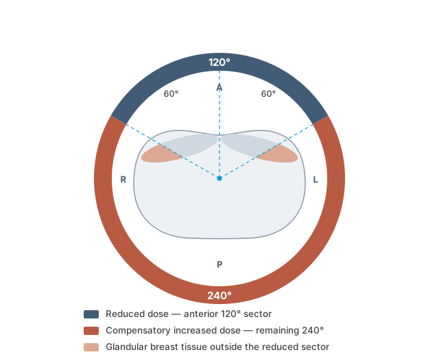
Figure 1: Schematic representation of organ-based tube current modulation in the breast region (120° sector, e.g., Siemens X-CARE; other systems use up to 180°). Own illustration.
For this dose reduction to take full effect, however, the tissue to be protected must lie within the intended range. In the supine position, this is frequently not the case: if the breast tissue shifts laterally, it may lie partly outside the reduced-dose zone. The tissue then benefits only partially, or not at all, from the intended protective effect. In large or ptotic breasts, this effect is even more pronounced.
Given the high radiosensitivity of glandular breast tissue, this issue is particularly relevant. If the actual position of the breasts is not sufficiently taken into account during the examination, the intended protection may not be achieved despite modern dose-reduction technology.
At the same time, radiation protection must not be considered in isolation. Reducing the dose too far can impair image quality and, in the worst case, make repeat scans necessary, which would increase the patient's overall exposure.
It is in this tension between image quality and radiation protection that careful positioning becomes decisive. A study illustrates how relevant this is in practice: in "Organ-based Tube Current Modulation: Are Women's Breasts Positioned in the Reduced-Dose Zone?", 99% of 532 patients examined in the supine position had at least part of the breast tissue within the increased-dose zone. This shows that the anatomical position of the breasts cannot be taken for granted when organ-based tube current modulation is applied.
3. When Small Measures Show a Big Effect: Practical Approaches
Positioning has to satisfy several requirements at once. It should be quick and easy to integrate into the existing workflow, respect the patient's privacy, and hold the breast as stably and reproducibly as possible. At the same time, it should help avoid image artifacts, unnecessary enlargement of the field of view, and an unfavorable dose distribution.
The technical parameters of the CT system also set clear limits. Depending on the manufacturer, the gantry bore is usually around 70 to 80 cm, and the diagnostically relevant field of view is generally limited to a maximum of 500 mm. For reliable assessment, both breasts should therefore be brought fully within this range at the positioning stage.
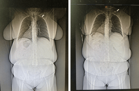
Figure 2: Comparison of a scout image without specific breast positioning (left) and with adequate breast positioning (right)
Patients also come to CT with very different body shapes, body weights, and breast sizes. In the supine position, breast tissue often shifts laterally, and this is frequently most pronounced in patients with obesity or with very large breasts. The more difficult it becomes to capture the entire breast within the field of view, the more important deliberate, patient-specific positioning becomes.
This matters not only for radiation protection but also diagnostically. For certain indications, such as melanoma, assessment of lymph nodes in the breast region, breast cancer, or possible incidental findings, complete coverage of the breast region can be decisive. In practice, three approaches in particular have emerged.
3.1 Bra: simple and quick, but not always sufficient
One obvious option is to let the patient keep her bra on, provided it is suitable for the examination. The advantage is clear: the measure is quick, requires no additional accessories, and can be more comfortable for the patient, because she needs to undress less. In addition, a bra can help keep the breast tissue closer to the thorax and reduce lateral displacement, which can allow a smaller field of view.
In practice, however, clear limitations become apparent. Not every bra is suitable for a CT examination. Metal underwires or fasteners can cause artifacts, particularly blooming artifacts. Bras also differ greatly in shape, material, and fit. Depending on the model, one breast may be well supported while the other continues to sink laterally. Despite the bra, gravity can therefore still cause breast tissue to come to rest laterally.
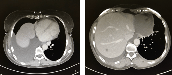
Figure 3: Visible blooming artifacts caused by a bra with metal components
3.2 Foam Positioners and Straps: more targeted positioning with greater control
Another option is positioning with aids such as foam positioners and straps. This allows the breast tissue to be positioned more precisely within the table area and the field of view. In the available practical examples, the breast could be positioned using foam positioners and a strap so that it was captured completely and with a smaller field of view.
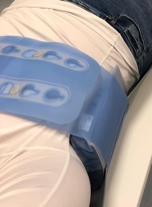
Figure 4: With foam positioner and strap (e.g. ProFoam and ProBelt).
The advantage of this approach lies in greater control. The positioning can be adapted to the individual anatomy and is less dependent on which bra the patient is wearing. This makes it potentially more reproducible. At the same time, the method requires a little more time and finesse. The aids must be placed correctly, without creating pressure points or placing unnecessary strain on the patient. In addition, the patient's privacy requires particular attention with this method.
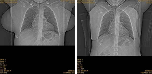
Figure 5: Left without positioning aids, right with foam positioner and strap (e.g. ProFoam and ProBelt). On the right, well positioned on the table and optimally captured with an FOV of 330.
3.3 WRAP-AROUND STRAPS: stable positioning for challenging cases
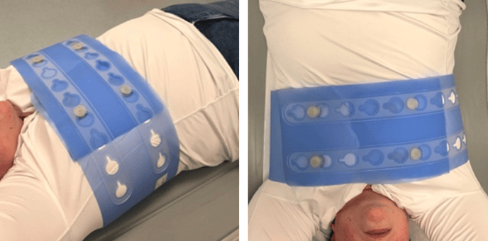
Figure 6: Examples of wrap-around strap application (ProBelt Wrap)
A wrap-around strap can be helpful when the breast tissue falls markedly laterally or when more stable, bilateral guidance is needed. The aim is not strong compression but gentle, even stabilization of the breasts closer to the thorax. Particularly in large or ptotic breasts, this can help limit the field of view and keep the breast within the relevant examination range.
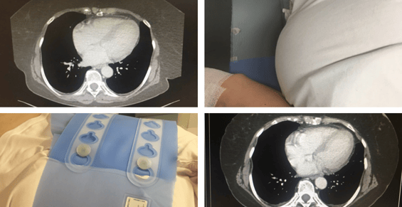
Figure 7: Comparison of positioning with and without a wrap-around strap (ProBelt Wrap)
Compared with a bra, the wrap-around strap allows more standardized positioning, because the CT team can actively control the position. It does, however, add a step to the workflow. Equally important is informing the patient clearly and carrying out the measure respectfully. The strap must not constrict, create skin folds, or cause painful pressure points.
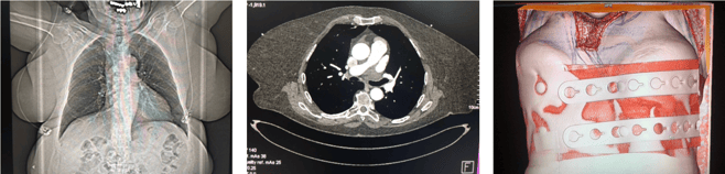
Figure 8: Left without positioning aids, centre with wrap-around strap (e.g. ProBelt Wrap 200); right in 3D reconstruction. On the left, despite an FOV of 500 the entire breast is not captured; centre/right the entire breast is captured.
Targeted positioning can also affect dose values in practice, as the following example shows:
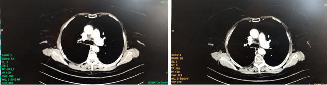
Figure 9: 368 mAs, image right: 279 mAs
4. Practical Conclusion
There is no single correct positioning for all patients. Which aid is appropriate depends on anatomy, clinical indication, and workflow. The following overview summarizes the key differences.
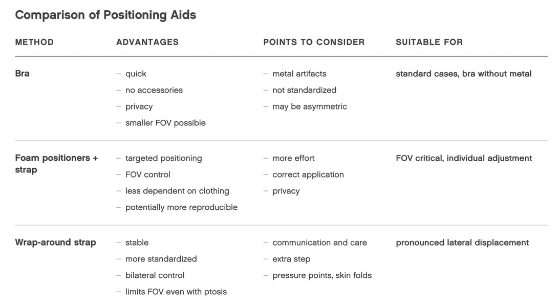
Figure 10: Comparison of Positioning Aids. Own illustration.
Regardless of the method chosen, a deliberate check before the scan is decisive: Does the breast tissue lie within the relevant range? Is the field of view as small as possible and as large as necessary? Is there any metal, skin folding, or asymmetric displacement? This brief check can help support image quality and radiation protection without unnecessarily complicating the CT workflow.
5. Conclusion: Deliberate Positioning as Part of Good CT Practice
CT examinations in patients with obesity make clear that standard technical solutions in radiological practice increasingly reach their limits. Large and ptotic breasts in particular are a relevant and, until now, often underestimated factor. This reveals a central problem: even modern technologies such as organ-based tube current modulation can only deliver their full protective effect if anatomical conditions are taken into account in practice. In these cases, the position of the breast largely determines whether the dose reduction actually reaches the tissue it is meant to protect.
Given the high and still rising number of patients with obesity, relying on standardized protocols alone is not sufficient. Instead, CT imaging calls for a deliberate, patient-specific approach in which positioning and individual anatomy are actively taken into account. This also reflects the thinking behind the ALARA principle, as low as reasonably achievable: not the lowest possible dose at any cost, but as little as possible and as much as necessary to achieve diagnostic image quality. A dose reduced too far can, in the worst case, make repeat scans necessary and thereby increase overall exposure.
Even simple measures can make a decisive difference here. A deliberate check of breast position, suitable positioning aids, or individually adapted positioning can help bring image quality, radiation protection, and workflow into better balance.
References
Bundesamt für Strahlenschutz. (n.d.). Grundsätze des Strahlenschutzes. https://www.bfs.de/DE/themen/ion/strahlenschutz/einfuehrung/grundsaetze/grundsaetze_node.html
Deutsche Adipositas-Gesellschaft. (n.d.). Definition von Übergewicht und Adipositas. https://adipositas-gesellschaft.de/ueber-adipositas/definition-von-adipositas/
Roth, J. (2004). Wie wirken sich ionisierende Strahlen auf den Menschen aus? Fachverband für Strahlenschutz e. V. https://www.fs-ev.org/fileadmin/user_upload/80_FAQs/strahlung_und_dosis/faq_frage_102.html
Krieger, H. (2007). Grundlagen der Strahlungsphysik und des Strahlenschutzes (2., überarb. und erw. Aufl.). Vieweg+Teubner. https://doi.org/10.1007/978-3-8351-9128-0
Robert Koch-Institut. (2024). Themenschwerpunkt: Übergewicht und Adipositas. https://www.rki.de/DE/Themen/Nichtuebertragbare-Krankheiten/Koerperliche-Gesundheit/Adipositas-und-Uebergewicht/themenschwerpunkt-adipositas.html
Taylor, S., Litmanovich, D. E., Shahrzad, M., Bankier, A. A., Gevenois, P. A., & Tack, D. (2015). Organ-based tube current modulation: Are women’s breasts positioned in the reduced-dose zone? Radiology, 274(1), 260–266. https://doi.org/10.1148/radiol.14140694
Über-Gewicht. (n.d.). Adipositas: Definition, Behandlung & Folgen der Erkrankung. https://www.ueber-gewicht.de/adipositas.html
Wirth, A., & Hauner, H. (Hrsg.). (2008). Adipositas: Ätiologie, Folgekrankheiten, Diagnostik, Therapie (3. Aufl.). Springer Medizin.