In radiology and especially in MRI examinations, motion artifacts play an important role. Uncooperative patients, challenging examination positions or too little time for correct patient positioning can lead to motion artifacts. These motion artifacts have a major impact on image quality, making it difficult to evaluate the results and can even lead to misdiagnosis. Therefore, motion artifacts are a frequent cause that individual sequences or even entire examinations must be repeated. A circumstance that has an impact on many areas of everyday clinical practice. Patient satisfaction decreases, the duration of MRI’s increases, employees are more stressed and the costs for examinations increase.
- How do motion artifacts occur?
- In which examinations or patients do motion artifacts occur?
- What influence do motion artifacts have?
- How can motion artifacts be avoided?
How do motion artifacts occur?
Motion artifacts are image disturbances that can occur in all imaging procedures, such as CT and MRI examinations. Motion artifacts are among the most common artifacts along with metal artifacts, annefacts, phase wrap and line artifacts. Patients should remain extremely still during the examination, as any involuntary movement of the patient, no matter how small, has a great impact on the image quality and consequently on the examination results. Depending on the examination, an average MRI takes up to 45 minutes, a period during which it is very difficult for any patient not to move. However, especially patients of advanced age, with neurodegenerative diseases, children or injured persons have problems to lie motionless for such a long time. Even the smallest movements, such as compensatory movements to lie down a bit more comfortably, sneezing, coughing, scratching, but also the heartbeat, breathing and swallowing can have serious consequences for the image quality.
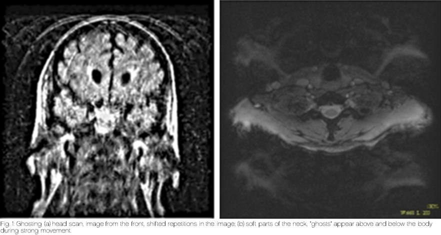
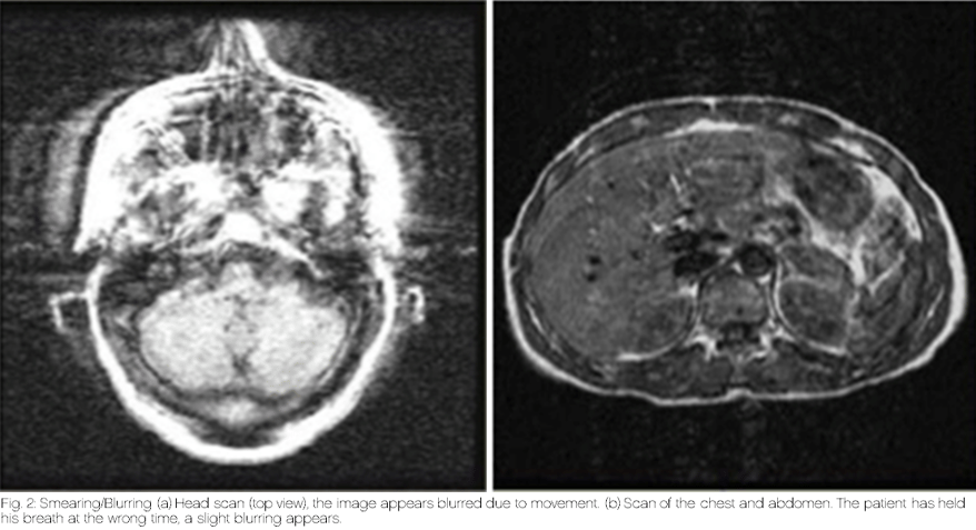
The most common motion artifacts include ghosting and smearing or blurring artifacts.
Ghosting causes “shifted repetitions” in the image and "ghosts" appear next to, above or below the body.
Smearing, or blurring, makes the image appear blurry or slightly out of focus.
In which examinations or patients do motion artifacts occur?
When asked this question, one is probably most likely to think of examinations in uncomfortable positions, which take a long time and are performed on rather uncooperative patients or children. One might think that there are fewer motion artifacts in outpatient practices because patients are often more cooperative and mobile. They show up for their appointment, lie down in the MRI, and then leave. These patients in particular give the appearance that they can lie still for 15 - 45 minutes without any problems. Unfortunately, this is not always the case.
The study of the authors Andre et al., which dealt with movements in MRI, showed that repeated or aborted sequences occur more frequently in the course of an examination - and this completely independently of the cooperation or "frailty" of the patients. In the first 5 minutes there is a relatively high number of repeated or aborted sequences, often due to patients who do not tolerate an MRI examination in principle, e.g., due to claustrophobia. If an examination lasts longer than 11 minutes, repetition sequences and aborted sequences increase more and more. The restlessness increases, the situation becomes increasingly uncomfortable and spontaneous convulsions occur more frequently.
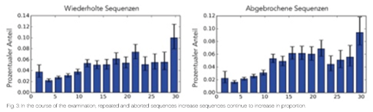
These results show that the issue of motion artifacts can only be related to a limited extent to the patient; rather, repetition sequences occur more frequently with increasing examination duration.
What influence do motion artifacts have?
Even the smallest movements in the MRI lead to a reduction in image quality and thus to losses in the evaluability of the data. Therefore, in such cases, repetition sequences are often required, which take additional time. And time is money, as we all know.
But not only the costs, but also the waiting time of the patients increase, the overtime piles up, the stress increases which again decreases the satisfaction of patients and staff.
The time available for adequate positioning and immobilization of the next patient decreases and movement artifacts tend to increase as a result. A vicious circle!
Of the limitations mentioned, patient satisfaction is one criterion that is directly measurable and has a direct impact on work morale and the workplace environment. Based on a survey of 6500 patients conducted by Brown University's Warren Alpert Medical School, wait times have a tremendous impact on patient satisfaction (Dibble et al., 2017).
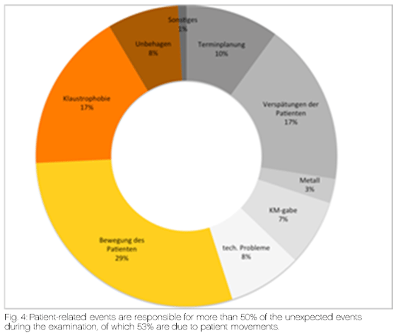
Based on this, according to a recent study from Emory University in Atlanta, about 17% of all scans are affected in some way by unexpected events, causing delays in the daily clinical routine (Sadigh et al., 2017). The majority of these events were caused by patient-related incidents during the scan (55%), with patient movement leading the way, followed by claustrophobia and patient discomfort.
According to this study, movements in the MRI have a direct impact on waiting times and patient satisfaction.
But it is not only patient satisfaction that is declining. Nowadays, an institution and its investments are increasingly evaluated on the basis of profitability. How can I generate a higher flow of patients in less time, with as few resources as possible, and thus increase profit?
A research group at the University of Washington has taken a closer look at this issue (Andre et al., 2015). They analyzed 192 MRI examinations and 1238 sequences contained therein.
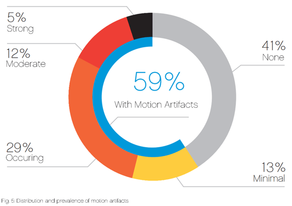
They found that severe to moderate motion artifacts occurred in approximately 17% of the sequences (203), making radiological interpretation impossible or at least extremely difficult. Mild motion artifacts occurred in another 42.3% (524) of the sequences. According to their statement, motion artifacts of varying severity were found in nearly 60% of the sequences.
 Repeats of individual sequences occurred in approximately every 5th MRI. Using standard national health insurance rates, they calculated the impact of these repeat sequences on the annual budget. According to the study, one minute of scanning costs just under $10. An average sequence takes about 4 min and 10 seconds, so it costs about $40. That doesn't sound like much at first. But if you multiply this by the number of repetitions in a week and extrapolate it to a year, you come up with an amount of $115,000 per year per scanner.
But not only the costs, but also the waiting time of the patients increase the overtime piles up, the stress increases and the satisfaction of the patients and the staff decreases.
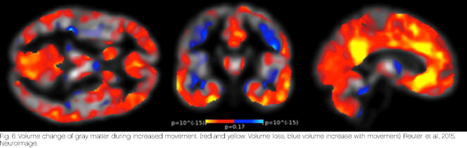
Researchers at Harvard University were able to show that patients' movements during the examination could cause the interpretation of the MRI results in the wrong direction (Reuter et al., 2015). For example, in Fig. 5: Distribution and prevalence of motion artifacts, certain artifact-rich structures may appear more conspicuous or less conspicuous than they actually are. Using the example of neurodegenerative diseases, such as Parkinson's or Alzheimer's disease, they found that the greater the head movements during the MR examination, the more the gray matter volume is reduced and the cortex appears narrower. There may be a shifted, incorrect assessment of quantitative brain analysis. Atrophy of the gray matter presents itself in an MR image as blurred areas, which are very easily mistaken for blurring artifacts due to motion. These motion artifacts can lead to misdiagnosis or misinterpretation of the severity of the disease, especially in neurodegenerative diseases. Therefore, it is important to clearly exclude all images that show deficiencies in the visual quality control as well as images that are provided with warnings by the system. This is especially important for patients who find it difficult to remain still for the entire duration of the examination. This poses a significant problem for patients who find it difficult to lie still for the entire duration of the examination, as the data set is extremely minimized.
How can motion artifacts be avoided?
To reduce the effects of motion artifacts described above, there are different approaches.
One possibility is to use software programs that identify motion artifacts during data processing and correct them afterwards using certain algorithms. However, since the focus should be on avoiding motion artifacts, this will not be discussed in more detail here.
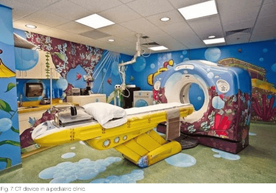
In contrast, patient comfort systems are an approach to distract patients from the often unfamiliar situation, to calm them down, and thus to avoid motion artifacts from the very beginning. On the one hand, the examination environment is designed to be as pleasant as possible, for example by means of relaxing room lighting, so that the patient's general well-being and tolerance for the examination increases. For children even, entire "adventure worlds" are created, which are integrated into the MRI/CT and the room, so that the examination is transformed into a kind of game for them. Furthermore, so-called in-bore solutions are increasingly being used, whereby the patient is involved in the examination process while lying in the scanner and is continuously informed about the progress. In addition, the patient is offered an "entertainment program" with sound and images.
Nevertheless, sedation is still resorted today to uncooperative patients and especially in children to ensure a smooth and motionless procedure.
One of the most important approaches involves direct interaction with the patient. Proper explanation and calm communication prior to the examination are just as important as adequate, in other words stable and comfortable positioning of the patient. In the case of claustrophobic patients, for example, a detailed explanation and shortened waiting time can be particularly helpful in order not to increase the patient's tension even further.
For adequate patient positioning, well-rehearsed workflows and the choice of the right positioning aids are essential. The aim of these is to ensure that the patient lies comfortably on the table, is positioned stable outside the coil and is comfortably fixed inside the coil. In this way, the examination results can be positively influenced, and the patient's comfort increased.
There are a variety of different systems, all of which fulfill their specific purpose and are often used in combination.
- What aspects should be considered when choosing the right storage aids?
- What modality must the positioning aid be compatible with (MR-safe, radiolucency, etc.)?
- In which situations is the positioning aid being used (inside coils, on the patient table as a support, etc.)?
- What goal is to be achieved during positioning (comfort, stability, achieving a specific position, etc.)?
- What special influences is the positioning aid exposed to (contamination, weight/force, germs, etc.)?
- How can permanently reliable hygiene be ensured, and which disinfectants can be used for this purpose?
One of these solutions for MRI are the so called MULTIPADs by Pearl Technology AG:
The MULTIPADs are inflatable systems and thus provide individual and good fixation within coils and trays. The positioning aids consist of 2 chambers, one of which is filled with polystyrene beads and the other can be filled with air using a hand pump. This allows the cushions to bridge different sized voids between the patient and the coils/shells. The pressure can be individually adjusted to the patient, allowing the ideal degree of fixation. Because of the beads, the entire surface of the cushion conforms to the patient's anatomical structures, ensuring even pressure distribution without pressure points (Haller et al., 2018).
MULTIPAD positioning aids are, MR safe and compatible with MRI equipment from all manufacturers. Thanks to an even and symmetrical pressure distribution a optimum level of comfort can be achieved even when stabilizing challenging patient groups. The universal applicability also reduces the variety of aids required and thus helps to optimize workflows. The MULTIPAD is easy to clean and therefore also has a positive contribution to patient safety and well-being.


CONCLUSION
It is hard to imagine what effects a small twitch at the wrong time can have. Thus, motion artifacts not only have an impact on the profitability of your radiology department, but also affect the satisfaction of your patients or make the interpretation of the data considerably more difficult. With relatively simple means, it is possible to prevent major effects. Stable and comfortable positioning of patients can simplify workflows, reduce costs and increase patient satisfaction.
Credits to: Angela Treis for providing this content
ABOUT US
Pearl Technology AG specializes in the development of innovative positioning aids in medicine, particularly in the fields of radiology and radiotherapy.
The patented technology, which originated in the research of Rheumatoid Arthiritis research using high-resolution pQCT systems, is characterized by high variability, uniform pressure distribution and adaptive fixation. It was developed in collaboration with ETH Zurich and renowned university hospitals.
As an ISO 13485 certified medical technology company, Pearl Technology AG relies on biocompatible, easy-to-clean materials and high-quality workmanship in Switzerland.
In addition to the development of a wide range of support aids, some products have already been realized as customer-specific solutions together with international equipment manufacturers.