
By Katrin Hägele – Your Expert in Advanced Imaging Systems
(Translated from German)
Cardiac computed tomography (commonly called Cardio-CT or coronary CT angiography, CCTA) is a highly advanced method for visualizing the coronary arteries and heart structures with precision. To achieve optimal results, it is crucial to minimize possible sources of error. Even small missteps, minor deviations, or isolated mistakes can significantly degrade—or even invalidate—the diagnostic outcome.
Thorough preparation, clear patient communication, meticulous execution, and final quality control of the images are all indispensable. Below you’ll find key points to pay attention to, regardless of the specific CT system in use.
Contents
Patient Preparation – The Key to Ideal Image Quality
Important Notes on Beta-Blocker Administration for Cardio-CT
Patient Positioning and ECG – Ensuring Clean Signals
Tips for Optimal Electrode Preparation
Breath-Hold Command and Scan Planning
Other Critical Details That Make a Difference
Conclusion
Sources
Patient Preparation – The Key to Ideal Image Quality
Careful patient preparation is critical for a successful CCTA. Be sure to observe the following:
-
Avoid caffeine and nicotine: For at least 12 hours before the scan, patients should avoid coffee, coke, black tea, green tea, energy drinks, etc. Caffeine stimulates the heart and increases the heart rate, which can lead to motion artifacts that impede evaluation of the coronary arteries. Nicotine likewise affects heart rate and blood pressure—both altering image quality. (Per ESC Guidelines 2024)
-
Avoid exertion and stress: Activities like climbing stairs just before the exam should be avoided. Anxiety or agitation in the waiting or scan room can also raise heart rate. A calm environment is essential—discussions or conflicts should not take place in the patient’s presence.
-
Blood pressure–lowering medications (Beta-blockers): These are used prior to the scan to reduce the heart rate to ~60 bpm and to stabilize it. A lower and more stable heart rate reduces motion artifacts, allows for shorter and more precise scan times, and leads to better coronary artery visualization. It also prolongs the diastole, giving a more motion-free window for imaging, and thereby helps reduce radiation dose.
Target: 60 bpm. This enables motion-minimized imaging between 60–70 % of the cardiac cycle, with minimal motion in all three coronary arteries.

Target: 75 BPM. Unlike lower heart rates, there is no low-movement phase here.

Achenbach S et al. (2000), Lu B et al. (2001), Husmann L et al. (2007)
Positioning and ECG – For Uninterrupted Signals
Accurate positioning and reliable ECG signals are essential in CCTA. Key recommendations:- Raise the arms: Arms should be elevated above the head to avoid thoracic artifacts. If arms remain in the scan field, X-ray attenuation may cause artifacts, compromise coronary assessment, and increase radiation dose. Arms must be raised before applying ECG electrodes to avoid skin folds or movement that would impair electrode contact.
- Comfortable positioning: The patient must be in a position they can hold stably, since even tiny movements may degrade ECG signals or create image artifacts. Use supportive aids if necessary, and confirm with the patient that the posture is maintainable for several minutes.
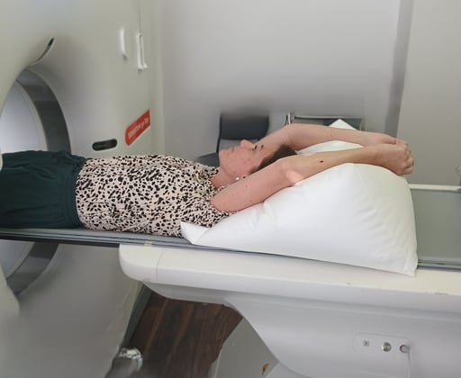
Photo: Hägele
Iso-center the heart: Standard practice is to center the heart in the imaging volume. Some vendors recommend a slight right shift to center the heart more precisely.

Image source: GE HealthCare GmbH
- Impedance control: Use the CT vendor’s recommended electrodes (within expiration dates). Ensure properly applied electrodes, and (if needed) shave or clean the skin to improve contact. Perform impedance checks: high impedance indicates poor signal quality, which may severely affect ECG-triggering.
In modern CTs, impedance is displayed (e.g. via “traffic light” color codes: red = invalid, yellow = acceptable, green = optimal). If impedance is poor, reapply electrodes. - Clean ECG leads: The ECG is the trigger source for image acquisition. Faulty signals can lead to incorrect triggering and blurred images. The software often highlights the detected R-wave; if the R-wave is ambiguous, triggering fails.

Some manufacturers use a traffic light system for impedance control:
Red = invalid
Yellow = acceptable
Green = optimal
Breath-Hold Command & Scan Planning
Additional aspects critical to image quality:
- Practice the breath-hold command: Consistent breath-holding is crucial to reduce motion artifacts — suboptimal breathing is a major cause of degraded scans. Before scanning, rehearse the breathing command until reproducible technique is achieved. Don’t hesitate to repeat as needed.
The patient should be able to hold breath for ~12 seconds, inhale consistently each time, avoid a Valsalva maneuver (i.e. don’t forcibly exhale through the nose), and avoid exhaling mid-hold. About 5 seconds after inhalation, heart rate tends to drop—this is often the optimal window for scanning.

Bildquelle: GE HealthCare GmbH
- Calcium scoring: A native (non-contrast) calcium scan helps quantify coronary calcification. This plays a key role in risk stratification and in planning further diagnostics or therapy. In cases of heavy calcification, a CCTA may be of limited diagnostic value and could be omitted. (Achenbach et al. 2012)
- Scan planning: Planning ensures all relevant coronary segments are captured while minimizing radiation dose. Use the native scan as a guide.
- Ensure the most superior coronary artery origin is fully within the scan range.
- Account for variants—e.g. vessels may arch upward or bend (“kinking”)—so include coverage for about 5 slices proximal to the first vessel (e.g. LAD) and 5 slices distal to the heart base, roughly 1–1.5 cm extra.
- Some post-processing software (vendor-dependent) may require standardized scan parameters: fixed reconstruction zone, slice thickness, tube voltage settings (e.g. 25 cm, 2.5 mm slices, 120 kV).


Image Source: GE HealthCare GmbH
Other Important Points: Details That Make the Difference
- Nitro spray: The sublingual administration of nitrates shortly before the scan dilates the coronary arteries and enhances depiction of small vascular segments, which aids in the evaluation of stenoses and anatomical variations. Be mindful of contraindications: in patients taking PDE-5 inhibitors (e.g. sildenafil, Viagra®) or in existing hypotension, nitroglycerin may further lower blood pressure and precipitate circulatory collapse. The exact timing of administration is critical to image quality. Ideally, the scan should take place 3 to 5 minutes after application when the effect is maximal.
Application of Nitrolingual® Pumpspray
⏱️ Time to onset: 1–2 minutes
⏱️ Maximum effect: 3–5 minutes
⏱️ Duration: 20–30 minutes
(Source: Fachinfo.de, rd-factsheets.de)
- Contrast volume & flow rate: The contrast agent volume and flow rate should be weight-adapted and depend on the CT system used. For a standard patient, a typical protocol uses 60 mL of a high-concentration contrast agent (320 or 350 mg iodine per mL) at a flow rate of 4.5 mL/s — generally sufficient to opacify the coronary arteries and reliably detect stenoses. For better depiction of the interventricular septum, a higher contrast volume may be needed; other indications may require different values. Manufacturer and application specialist recommendations can provide helpful orientation.
In general:
"The faster and more modern the CT scanner, the shorter and smaller the contrast bolus needed."
💉 Slower CTs (e.g. 64-slice) require more contrast and longer bolus because imaging spans several heartbeats.
💉 Fast CTs (e.g. 320-slice, high-pitch) can operate with a smaller, shorter bolus because they often capture the heart within a single cardiac cycle.
- Determining optimal scan timing via test bolus or bolus tracking: Two proven techniques are used to detect the optimal scan start time. Both rely on monitoring the contrast progression and start with planning a test slice at the level of the tracheal bifurcation.
|
Test bolus method |
Bolus tracking method |
|
A small contrast amount (e.g. 3 mL) is injected at the planned flow rate, followed by 20 mL of saline. Single CT slices are then taken to determine when the contrast reaches the target region (region of interest, ROI). From this, the delay (time between injection and optimal start) can be computed. |
A ROI is placed in the aorta (and sometimes in the central pulmonary arteries). The scan is triggered automatically once contrast arrives in the ROI and a preset Hounsfield Unit (HU) threshold is reached. Bolus tracking is more reliable, reproducible, and less user-dependent than the test bolus method, and less susceptible to unexpected events (e.g. extrasystoles). |
Bolus tracking is more reliable, reproducible, and less user-dependent than the test bolus method, and less susceptible to unexpected events (e.g. extrasystoles).
- Quality control: Immediately after the scan, image quality should be assessed. Early quality control enables detection of errors and, if needed, repeat scanning or corrective postprocessing — avoiding unnecessary repeat scans later.

![]()
Example of a sufficiently good examination. Good contrast in the coronary arteries. The heart is displayed without steps.

Another example after 3D post-processing (obligatory vessel and volume display): The coronary vessels are clearly and continuously displayed. Image source: Rolf et al. 2023
Pay particular attention to blurring. A stepped representation of the liver and diaphragm or sternum indicates breathing artifacts. This occurs primarily with mid-range CT scanners, such as 64-slice devices, which cannot capture the heart within a single heartbeat.


Example of a breathing artifact: A step in the sternum or diaphragm indicates that the patient has breathed. Image source: GE HealthCare GmbH
Conclusion
Cardiac CT is a highly sensitive procedure that only fulfills its full potential when all details are correct — from patient preparation and positioning to image post-review. Even minor deviations can significantly weaken diagnostic value. A calm environment, stable heart rate, precise ECG signals, and strict quality control are therefore critical success factors.
Adhering to these points consistently lays the foundation for reliable results and significantly helps to avoid repeat examinations, unnecessary radiation exposure, and misdiagnoses — all for the benefit of patient safety and efficient workflow within the CT team.
References & Useful Links
Achenbach et al., 2000
Achenbach, S., Ropers, D., et al. (2000). In-plane coronary arterial motion velocity: measurement with electron-beam CT. Radiology, 216, 457–466.
Achenbach et al., 2012
Achenbach, S., Barkhausen, J., Beer, M., u. a. (2012). Konsensusempfehlungen der DRG/DGK/DGPK zum Einsatz der Herzbildgebung mit CT und MRT. Kardiologe, 6, 105–125. Link
DGK, 2023
Deutsche Gesellschaft für Kardiologie – Herz- und Kreislaufforschung e.V. (DGK) (2023). Positionspapier: Diagnostik der koronaren Herzkrankheit mittels CT und MRT. DGK-Leitlinien, 11 S. Link
ESC Guideline 2024
Vrints, C.; Task Force for the Management of Chronic Coronary Syndromes der European Society of Cardiology; Endorsed by the European Association for Cardio-Thoracic Surgery (2024): ESC Guidelines für das Management des chronischen Koronarsyndroms. European Heart Journal, Band 45, Heft 36, S. 3415-3537. DOI: 10.1093/eurheartj/ehae177
Fachinfo Nitrolingual® Pumpspray
G. Pohl-Boskamp GmbH & Co. KG. Fachinformation Nitrolingual® Spray. In: Fachinfo.de [PDF]. Abrufbar unter: https://www.fachinfo.de/fi/pdf/020573 (Stand zuletzt geprüft) Fachinfo
If you need detailed page numbers: this product information contains information on application, dosage, and side effects (e.g., onset of action, frequency). Product information
GE HealthCare GmbH, Marketing-Department Germany (2025). With kind permission.
Husmann et al., 2007
Husmann, L., et al. (2007). Coronary artery motion and cardiac phases: dependency on heart rate—implications for CT image reconstruction. European Radiology, 17, 2010–2018.
Lu et al., 2001
Lu, B., et al. (2001). Effects of window and threshold levels on the accuracy of three-dimensional rendering techniques in coronary artery electron-beam CT angiography. Radiology, 218, 703–711.
Rettungsdienst Factsheets (Glyceroltrinitrat)
Rettungsdienst Factsheets. Glyceroltrinitrat. Abrufbar unter: https://rd-factsheets.de/fs/glyceroltrinitrat-2/ (Stand zuletzt geprüft) RD Factsheets
Rolf et al., 2023
Rolf, A., Schmermund, A., Hell, M. M., u. a. (2023). Qualitätskriterien für die Erbringung kardialer CT-Leistungen. Kardiologie, 17(2), 81–94. https://doi.org/10.1007/s12181-023-00599-z