 Implementing a photon-counting CT represents far more than a technological upgrade—it marks a genuine transformation in everyday radiology practice.
This shift was felt especially intensely at Zuger Kantonsspital: as the first hospital in Switzerland, it was granted permission to use the NAEOTOM Alpha.Pro in clinical routine.
But even before the first patient lay on the table, another issue was already at the forefront:
How will our work change—and how will the team feel about it?
After four months of clinical use, Sari Tillmann, CT Key User at Zuger Kantonsspital, shares her experiences.
Inhalt
1. Photon-Counting – What is the Technology Behind it?
2. TransfoRmation and Team Reactions
3. Core Features of the New Technology
4. Important Considerations for Technologists
5. A Key Feature: Spectral Post-Processing
6. Clinical Examples from the First Months
7.
Conclusion
Sources
A Technical Quantum Leap—and a Human Story
When it became clear that the incoming scanner would not be a traditional high-end CT but a photon-counting system, the story took an unexpected turn.
What followed was a mix of curiosity, pride, and healthy skepticism—reflecting what many departments experience when cutting-edge technology suddenly becomes part of daily operations.
These team reactions highlight what technological innovation truly means in clinical reality.
1. Photon-Counting – What Is the Technology Behind It?
To this day, most CT scanners operate with so-called energy-integrating detectors. Each incoming X-ray photon first generates a light signal, which is then read electronically. This classical setup is robust, but it comes with physical limitations.
One crucial factor is the reaction speed of scintillators:
Even modern materials require around 10,000 nanoseconds to generate a light signal. Yet in that same timeframe, roughly 30,000 photons strike a detector element. Mathematically, that leaves only about 8 nanoseconds per photon—far too little to detect each individually.
The result:
The detector produces a summed, energy-weighted signal in which subtle differences between photon energies are lost.
Photon-counting detectors work fundamentally differently. They bypass the light-conversion step entirely and convert each photon directly into electrical charge. The amount of charge corresponds to photon energy—lower energy produces less charge, higher energy more.
The advantage is clear:
Every photon is counted individually and simultaneously assigned its energy.
This inherently generates spectral information—without dedicated dual-energy modes or additional hardware.
The technology enables more precise, differentiated, and efficient CT imaging—laying the foundation for the next generation of computed tomography.
2. Transformation and Team Reactions
Before it was decided that a photon-counting CT would be installed at Zuger Kantonsspital, the original plan was to acquire a SOMATOM Force. Preparations were already underway when the renovation was suddenly halted. New negotiations began, and even our team of technologists initially did not know what was happening behind the scenes. This phase was marked by anticipation, open questions, and a tangible sense of suspense.
When the final decision was announced—that we would be receiving a photon-counting CT—it was a true personal highlight for me. It was the moment I realized that we were taking a major technological leap forward.
As CT Key User, it mattered to me how the entire team perceived this change. Feedback was overwhelmingly positive: pride, excitement, and genuine enthusiasm shaped many discussions. Comments like “We are helping shape the future” or “New technology motivates us” demonstrated how strongly the decision resonated within the team.
At the same time, some colleagues voiced skepticism. They questioned whether the new technology would truly offer measurable benefits for patients, how significantly everyday work might change, or what further training would be required. Others found the term “photon counting” hard to grasp and worried that an entirely new system might introduce uncertainty.
However, this mix of excitement and hesitation quickly faded once the many advantages became evident in clinical routine. The excellent image quality, flexible scan modes, and noticeable workflow improvements strengthened the team’s confidence, turning initial doubts into real enthusiasm.
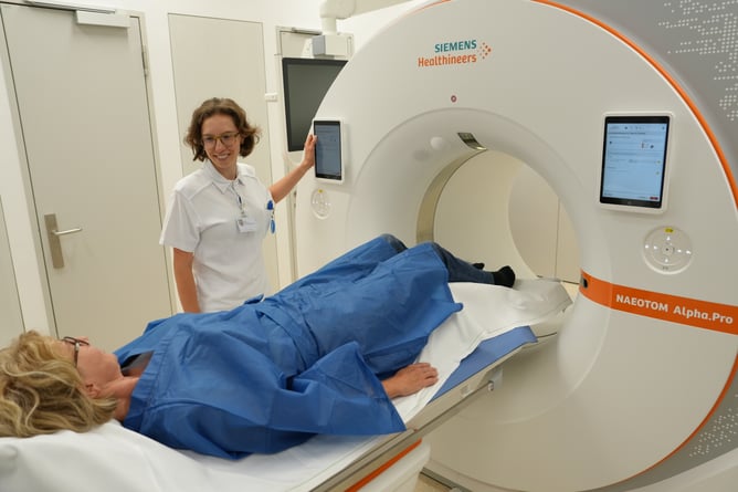
Figure 1: Sari Tillmann examining a patient.
In the end, it became clear:
Introducing the new system was not only a technical milestone—it also enhanced our emotional connection to our work. A shared step into a new era of imaging.
3. Core Features of the New Technology
As soon as clinical operations began, it became apparent why photon-counting technology received such strong support from the team. Many theoretical advantages became immediately visible in daily practice, confirming that the decision had been the right one.
Flexible Scan Modes – Tailored to Every Clinical Need
The four main modes (Quantum, Quantum.Plus, Quantum.Tin, and Quantum.Peak) allow precise adjustment to the clinical indication. This enables targeted balancing of image quality, speed, and radiation dose—particularly helpful for complex cases or patients with limited tolerance.
Extensive Reconstruction Options
Spectral raw data provide a wide range of image types simultaneously. This simplifies interpretation, improves tissue differentiation, and reduces artifacts—without requiring additional scan phases.
Customizable keV Levels
Reconstruction can be performed at any desired energy level (e.g., 40, 70, or 120 keV). This allows radiologists to optimize image appearance based on contrast distribution, scan phase, or clinical question before sending images to PACS.
Reduced Radiation Dose and Contrast Medium
In routine clinical use, dose reductions of up to 50% have been achieved—while delivering equal or better image quality. In one comparison between the SOMATOM X.ceed and the Alpha.Pro, radiation dose was reduced by 49% in a patient with a BMI of 45, while diagnostic image quality clearly improved.
Contrast medium consumption also benefits from spectral technology. Depending on the exam, reductions of up to 50% are possible; on average, reductions of around 30–35% were observed compared to the SOMATOM X.ceed. This not only improves patient safety but is especially beneficial for patients with impaired kidney function.
Virtual Non-Contrast (VNC) – Less Effort, Less Dose
Native scan? Often no longer necessary.
VNC reconstructs a native phase directly from the spectral dataset—saving time, radiation, and additional scan phases.
Patient-Oriented Acquisition – Flexible and Fast
Acquisition parameters can be adapted precisely to anatomy and clinical needs.
- Ultra High Resolution (UHR) mode: slices as thin as 0.2 mm for maximum detail.
- Flash mode: extremely fast scanning thanks to dual-source technology—ideal for cardiac CT, respiratory motion, or restless patients.
Expanded Diagnostic Spectrum
Thanks to thin slices and reduced metal artifacts, clinical questions previously difficult to answer can now be addressed more reliably—such as CTEPH, gout, bone marrow diagnostics, stents, or peripheral vascular occlusions.
Shorter Examinations
Not only Flash mode but also other scan modes—especially with tin filtration—shorten exam durations significantly. Compared with the X.ceed, many exams are now completed noticeably faster.
Energy-Efficient Operation
Eco-Power mode reduces energy consumption during low-activity periods—important because the scanner cannot be fully powered off.
Greater Diagnostic Confidence
Clearer spectral information makes it easier to detect, differentiate, and assess findings. Radiologists benefit from sharper structural detail and improved differential diagnosis.
4. Important Considerations for Technologists
Switching to a photon-counting CT introduces several particularities in daily work. Many became routine quickly, while others require more deliberate attention. Key lessons from the first months include:
Do Not Power the System Off Completely
The photon-counting detector must not be fully powered down. If the detector loses power, the system needs time to regain full image quality. Although scanning is possible before stabilization, image quality will be noticeably reduced. Proper team training is essential to ensure optimal performance.
Reconstruction Time and PACS Transfer
Despite some early concerns, photon-counting systems do not cause delays—provided the IT infrastructure is configured appropriately. With the Alpha.Pro, reconstruction time and PACS transfer remain at familiar levels.
Important preparation step:
The network should be tested with Siemens reference data and adjusted if necessary to ensure smooth data flow.
Unlocking the Full Potential
To fully benefit from the photon-counting CT, teams must be willing to question existing routines.
This is particularly relevant for:
- contrast protocols
- selection of reconstruction types
- usage of scan modes
The technology opens many new possibilities—but these can only be realized if technologists and key users actively explore and test them.
Familiar Interface: VB10 ≈ VB20
A major advantage during onboarding: the Alpha.Pro interface closely resembles previous versions. Staff familiar with VB10 need to learn only about 10–20% new content. For teams with dual-source experience, the learning effort is even lower.
Patient Positioning
Stable, precise positioning is especially critical with photon-counting CT, as scans are performed very quickly and even small movements can have visible effects. The head CT scan (spiral acquisition) is particularly sensitive to artifacts, making exact and secure head positioning essential.
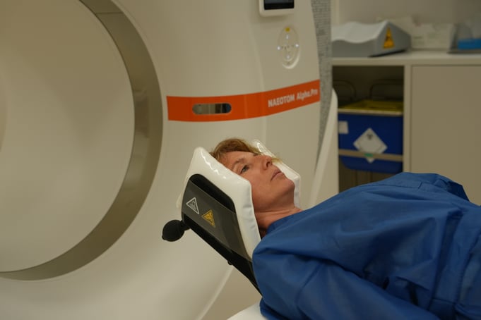
Figure 2: Head immobilization for stable and precise positioning in photon-counting CT.
A striking insight emerges when comparing conventional CT with photon-counting CT:
No new artifacts appear—the same artifacts simply become more visible.
The difference lies solely in how they are displayed. Many artifacts that were previously obscured by noise now appear more clearly. The “noise carpet” of older systems effectively concealed them. With direct detection and significantly lower electronic noise, photon-counting CT delivers very clear, low-noise images—making existing artifacts more apparent.
This increased clarity is a major diagnostic advantage, as finer structures become visible. At the same time, it underscores the importance of proper positioning. The more stable the head is positioned, the lower the risk that artifacts will appear or become more pronounced.
In summary:
The photon counter does not create new artifacts—it simply displays existing ones more accurately.
Therefore, careful and stable positioning is a crucial part of everyday operation with this new technology.
5. A Key Feature: Spectral Post-Processing
Spectral post-processing is one of the most powerful unique features of photon-counting CT. Because photons are measured directly, all spectral information is automatically available—without special scan modes or additional acquisitions. This opens the door to highly precise and differentiated image evaluation.
Here are the most important tools and reconstruction types:
Monoenergetic Plus
Images can be reconstructed freely between 40 and 190 keV.
- Low keV levels (e.g., 45 keV) increase contrast differences—for example in vessels, liver, or aorta.
- Higher energies help reduce noise and artifacts.
VNC – Virtual Non-Contrast
A native phase is reconstructed directly from spectral data.
This removes the need for an additional scan and benefits dose, workflow, and patient safety.
Iodine Map
Displays iodine distribution selectively, with bone structures visible for orientation.
It is the counterpart to VNC—but focused on contrast medium assessment.
Pure Calcium & Pure Lumen
- Pure Calcium: shows calcifications only—ideal for angiographic studies.
- Pure Lumen: displays only contrast-filled lumen while suppressing calcium.
Both significantly improve stenosis and residual lumen evaluation.
T3D Reconstruction
Simulates a conventional CT image from spectral data—useful for comparison studies or consistent reporting.
Lung Analysis
Iodine-based perfusion imaging enables functional assessment of the lungs—particularly valuable in cases of pulmonary embolism.
Gout Reconstruction
Available as VRT or MPR. Gout deposits are displayed in color or grayscale—comparable to dual-energy systems, but without specialized DE scan modes.
Bone Marrow
Color-coded visualization of bone marrow edema simplifies assessment of lesions and inflammatory changes.
Rho/Z
Optimized for late-phase contrast imaging and used to visualize myocardium, scars, or fibrosis. Comparable to late enhancement in cardiac MRI—now possible in CT thanks to high iodine contrast and high spatial resolution.
SSP Job
All spectral data from a patient are fully stored, allowing additional reconstructions and analyses later—also outside the scanner console.
Automation Without Extra Effort
A major advantage for technologists: all spectral reconstructions run automatically in the background and require no additional work at the scanner.
6. Clinical Examples from the First Months
The first weeks with the photon-counting CT demonstrated how strongly the new technology influences diagnostic practice. These examples come directly from clinical routine and show how image quality, spectral information, and fast scan modes expand diagnostic possibilities—often at significantly lower radiation dose.
Gout
The new gout imaging is highly appreciated by referring physicians because it provides clear and quantifiable visualization. Deposits are highlighted in green and automatically measured. This simplifies reporting and makes findings more understandable for patients.
Video 1: VRT, Spatial extent of gout tophi (Volumetry: ) Video 2: MPR, Cross-section of the knee
Cardiac CT
Stent patients can be assessed almost artifact-free. The lumen before, inside, and after the stent is clearly visible. Thanks to Flash mode, cardiac CT can be performed at very low doses (approx. DLP 50) with image quality that clearly surpasses the previous system.
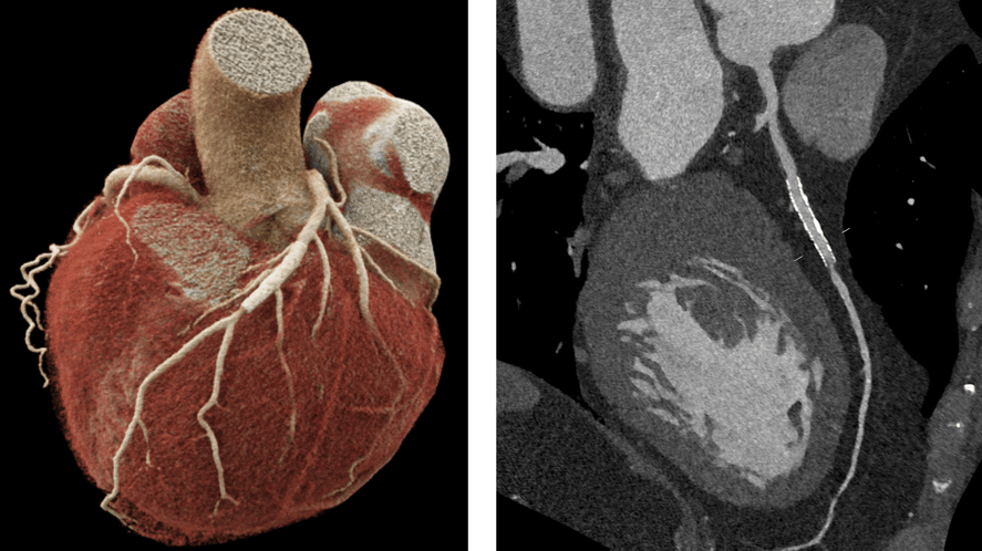
Figure 3: Modern cardiac imaging using photon-counting CT: Comparison between VRT (left) for anatomical overview and MPR slice (right).
Renal Hematoma
In a trauma CT, an unclear fluid collection near the kidney was observed. With VNC, a hematoma—not a urine leak—was confirmed clearly, without requiring an additional scan phase.
Low-Dose Lung
Compared with the previous AS+ scanner, the Alpha.Pro provides significantly sharper, more detailed images at the same dose (DLP 55). This becomes especially evident in zoomed views—without increasing dose.
Pediatric Lung
In a 9-year-old, a full inspiratory and expiratory lung scan was achieved at extremely low dose (DLP 12 mGy·cm). Despite minimal exposure, image quality remained high enough for a reliable assessment.
Breast Cancer
Spectral imaging supports therapy monitoring. In a breast cancer follow-up, the iodine map showed a clear decrease in contrast uptake in the residual tumor—indicating therapy response beyond size measurements.
Extremities with Metal
After a fall, a patient with a prior metacarpal fracture was suspected of having implant damage. Monoenergetic reconstruction at 150 keV showed the metal plates almost artifact-free. Both plates were clearly identified as broken. For soft tissue evaluation, lower keV levels would have been selected.
Perfusion Map
Perfusion maps support the detection of small pulmonary emboli that are easily missed in standard reconstructions. Color-coded images highlight perfusion deficits immediately, facilitating fast and intuitive reporting.
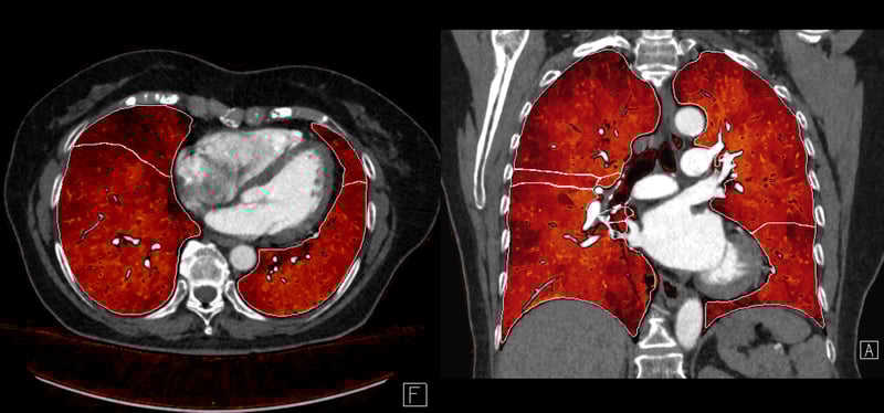
Figure 4: Color-coded visualization of pulmonary perfusion in two views.
7. Conclusion
Spectral imaging shows clear added value in everyday clinical practice—regardless of hospital size or examination scope. Patients benefit from higher image quality, reduced radiation exposure, and lower contrast medium usage. At the same time, the technology enables radiology teams to work more precisely, more efficiently, and overall more comfortably.
Photon-counting CT has the potential to transform modern imaging sustainably. Many tasks that were previously only partially feasible can now be performed with greater diagnostic confidence, higher resolution, and clearer differentiation. The development clearly points toward a future in which photon-counting detectors become standard—bringing lasting improvements to clinical workflows and patient experience.
Sources
Siemens Healthineers. NAEOTOM Alpha – Redefining computed tomography. Siemens Healthineers, Erlangen.
Siemens Healthineers. The technology behind photon-counting CT. Siemens Healthineers, Erlangen.
Siemens Healthineers. Spectral Imaging with Photon-Counting Technology. White Paper, Siemens Healthineers, Erlangen.
Zuger Kantonsspital AG, Baar. Own images / Material.